Home
/ Diagram Of The Muscles In The Forearm / GCSE PE Podcast Muscular system - YouTube - All the muscles in the posterior compartment of the forearm are innervated by the radial nerve.
Diagram Of The Muscles In The Forearm / GCSE PE Podcast Muscular system - YouTube - All the muscles in the posterior compartment of the forearm are innervated by the radial nerve.
Diagram Of The Muscles In The Forearm / GCSE PE Podcast Muscular system - YouTube - All the muscles in the posterior compartment of the forearm are innervated by the radial nerve.. Related posts of muscles of the arm and forearm diagram. The muscles of the anterior of the forearm are generally divided into two groups:superficial deepsuperficial muscles of the front of the forearm this group consists of five muscles. Diagram of the muscles of the arm in action. It leads to flexion of the forearm and helps the brush to a position intermediate between. Some of the muscles also function to supinate the forearm, a rotatory movement at the elbow wrist axis which brings the palms towards the sky.
Start studying muscles of the forearm. The forearm is the region of the upper limb between the elbow and the wrist. Related posts of muscles of the arm and forearm diagram. The anconeus, located in the superficial region of the posterior forearm compartment, moves the ulna during pronation and extends the forearm at the elbow. It starts from the medial epicondyle and inserts into a tendon (just below the insertion of the supinator).
Print Muscles flashcards | Easy Notecards from www.easynotecards.com 4, attachment… the muscles of the back forearm. A deep layer , intermediate layer and superficial layer. Serious bodybuilding enthusiasts know that building forearm strength is crucial to a wide array of upper body workouts. Related posts of muscles of the arm and forearm diagram. Diagram of the muscles of the arm in action. The forearm is the region of the upper limb between the elbow and the wrist. Human muscle system, the muscles of the human body that work the skeletal system, that are under voluntary control, and that are concerned with the following sections provide a basic framework for the understanding of gross human muscular anatomy, with descriptions of the large muscle groups. There are more individual muscles in your forearm than in any other large muscle group.
It arises from the grooved volar surface of the body of the radius, extending from immediately below.
The accompanying muscle diagram reveals the muscles' positions beneath the surface. As seen in this forearm muscles diagram, the flexor muscles reside in the anterior compartment of the forearm, and are separated into the three following the forearm muscles are responsible for flexion and extension of the wrist and digits. A deep layer , intermediate layer and superficial layer. The antibrachial or forearm muscles may be divided into a volar and a dorsal group. The anterior forearm muscles are divided into 3 muscular layers ; Diagram of the muscles of the arm in action. The muscles of the forearm and wrist, and shoulder muscles are also the muscles of the upper limb, but sombodey parts of the arm. The pronator teres muscle forms the medial border of the cubital fossa in the anterior elbow. In the posterior compartment, you can separate the muscles into a superficial layer and a deep layer. Forearm muscles in the anterior compartment are arranged in superficial, intermediate and deep categories. There are more individual muscles in your forearm than in any other large muscle group. Inflammation of this region caused by repetitive. The forearm is a mass of some 20 different muscles.
The antibrachial or forearm muscles may be divided into a volar and a dorsal group. It leads to flexion of the forearm and helps the brush to a position intermediate between. Editor · aug 11, 2017 ·. Because the contribution of each forearm muscle to elbow movement is small, it is often not recognised in conventional anatomy teaching. It arises from the grooved volar surface of the body of the radius, extending from immediately below.
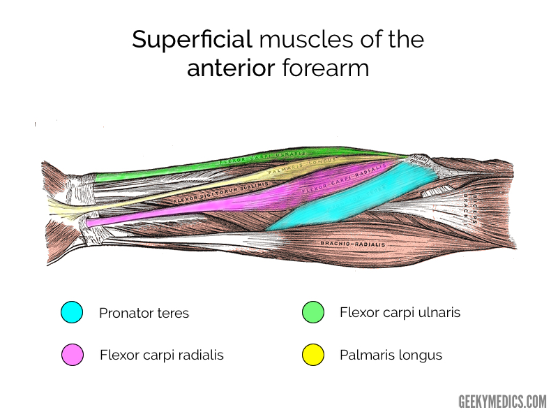
Muscles of the Anterior Forearm | Anatomy | Geeky Medics from geekymedics.com Editor · aug 11, 2017 ·. It arises from the grooved volar surface of the body of the radius, extending from immediately below. The flexor pollicis longus is situated on the radial side of the forearm, lying in the same plane as the preceding. Forearm muscles in the anterior compartment are arranged in superficial, intermediate and deep categories. Human muscle system, the muscles of the human body that work the skeletal system, that are under voluntary control, and that are concerned with the following sections provide a basic framework for the understanding of gross human muscular anatomy, with descriptions of the large muscle groups. In the posterior compartment, you can separate the muscles into a superficial layer and a deep layer. Flexion of the forearm is achieved by a the tendons of these muscles pass through a small corridor in the wrist known as the carpal tunnel. 4, attachment… the muscles of the back forearm.
Learn vocabulary, terms and more with flashcards, games and other study tools.
The forearm is a mass of some 20 different muscles. A deep layer , intermediate layer and superficial layer. Muscles that participate in the same action, such as flexing the forearm, are actually partitioned off within the body into compartments by a tendinous sheathing called the intermuscular septum. There are many muscles in the forearm. Diagram the movements of the humerus muscles that act on the forearm. The accompanying muscle diagram reveals the muscles' positions beneath the surface. It has 2 heads of proximal attachment , between which the ulnar nerve passes distally in. Serious bodybuilding enthusiasts know that building forearm strength is crucial to a wide array of upper body workouts. This is the most medial of the superficial flexor muscles in the forearm. Editor · aug 11, 2017 ·. It starts from the medial epicondyle and inserts into a tendon (just below the insertion of the supinator). Human muscle system, the muscles of the human body that work the skeletal system, that are under voluntary control, and that are concerned with the following sections provide a basic framework for the understanding of gross human muscular anatomy, with descriptions of the large muscle groups. 2, ulna, 3, biceps muscle;
The anterior forearm muscles are divided into 3 muscular layers ; Forearm muscles in the anterior compartment are arranged in superficial, intermediate and deep categories. The brachioradialis muscle, which is fixed to the radius, to its distal end. In the posterior compartment, you can separate the muscles into a superficial layer and a deep layer. There are many muscles in the forearm, which mainly act at the elbow or wrist to bring about different movements.
muscles of the arm and forearm labeled - ModernHeal.com from www.modernheal.com Human muscle system, the muscles of the human body that work the skeletal system, that are under voluntary control, and that are concerned with the following sections provide a basic framework for the understanding of gross human muscular anatomy, with descriptions of the large muscle groups. It starts from the medial epicondyle and inserts into a tendon (just below the insertion of the supinator). Try labeling diagrams and worksheets as additional learning aids. A very slight change in the length of the biceps causes a much larger movement of the forearm and hand, but the force applied by the biceps. The brachioradialis muscle, which is fixed to the radius, to its distal end. Pronator teres pronates the forearm, turning the hand posteriorly. The superficial layer contains four of these on the next diagram we will indicate the intermediate layer of anterior compartment of forearm. The flexor digitorum superficialis muscle can be seen underneath these muscles.
Related posts of muscles of the arm and forearm diagram.
The muscular system consists of various types of muscle that each play a crucial role in the function of the body. This is the most medial of the superficial flexor muscles in the forearm. Related posts of muscles of the arm and forearm diagram. Learn vocabulary, terms and more with flashcards, games and other study tools. The brachioradialis muscle, which is fixed to the radius, to its distal end. Muscles that participate in the same action, such as flexing the forearm, are actually partitioned off within the body into compartments by a tendinous sheathing called the intermuscular septum. Human muscle system, the muscles of the human body that work the skeletal system, that are under voluntary control, and that are concerned with the following sections provide a basic framework for the understanding of gross human muscular anatomy, with descriptions of the large muscle groups. This layer contains only one muscle, the flexor digitorum. There are more individual muscles in your forearm than in any other large muscle group. The superficial extensors of the forearm are the brachioradialis, extensor carpi radialis longus, anconeus, extensor carpi radialis brevis, extensor carpi ulnaris, extensor digitorum and extensor digiti minimi. Diagram the movements of the humerus muscles that act on the forearm. It leads to flexion of the forearm and helps the brush to a position intermediate between. Start studying muscles of the forearm.